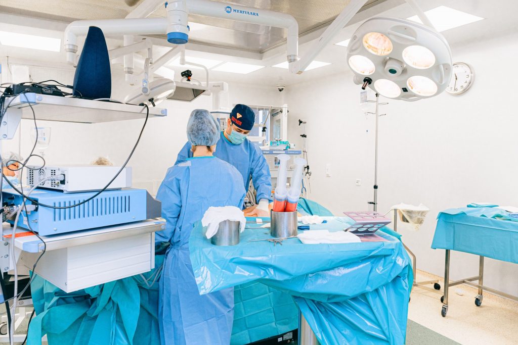
Image Guided Surgery (IGS) is the use of tracked surgical instruments. These instruments will vary according to what is being surgically accomplished and where in the body the surgery is taking place. Imagery being recorded prior to surgery and or actively during surgery will assist in making this process successful. The use of electromagnetic , cameras and ultrasonic are used in the IGS surgery system. The system helps to guide the surgeons into a safer and less intrusive surgery, where scars are barely visible or not visible at all.
Benefits of using Image Guided Surgery are reduced tissue trauma, as the surgeon may go directly to the correct anatomical specifications. The imagery shows exactly if there is swelling and organs that may be displaced slightly, so surgeons can avoid needing to alter anatomy but go safely around it as may be necessary. This is not only a safer benefit but a quicker procedure with minimal surprises. The surgeon will also have this imagery prior to the surgery. This gives the surgeon more knowledge of each person’s anatomy and can be better prepared with a more direct plan of operation.
- Safer
- Most are Less Invasive
- Quicker
- No scars or minimal scarring
For almost twenty years, IGS has used different image or tracking applications, but it was created for treatment of brain tumors initially. This process used a (CT) computed tomography, ( MRI) magnetic resonance imaging as well and other image capturing devices. Since then the ability to track during surgeries has greatly improved and insures very successful operation processes in the present day.
As of today the IGS systems use technical assistance in real time and during the actual operation. Such tracking assistance includes a hand held probe that gives surgeons the anatomical layout of the surgical area, during the surgery process. Then during the operation, the IGS displays the movement of the probe in real time. This opens up the use of the IGS for other surgeries as well.
Different surgeries possible using IGS are numerous now, however we see this technique used in sinus, organ repair, or removal, tumor removal that were once thought inoperable.
The IGS procedure is used when there is a need to remove or manage a problem within the human body. Most generally is it considered a standard procedure for areas such as; orthopedic, spine, cardiovascular and the otorhinolaryngology (ear, sinus and throat issues) and neurosurgery (surgeries to aid the nervous system throughout the body.
Image guided surgery information is recorded and can be used to gain process information and expose the need for more tools to assist. The technique is capable of handling several different approaches as needed. One example would be the fluorescent technology that helps to view the specific anatomical surgical area. Robotics can be an additional tool to help extract tissue or do implants, like joints or electrodes.
The IGS system allows the surgeon to visualize and navigate through the human body, in different ways. The use of different tools helps expedite the surgery process and make the surgery gain ground in otherwise unknown possibilities. There are fewer limits to what can be accomplished because of the increasing knowledge gained by using the IGS techniques and tools.
Surgeries that were considered impossible are quite possible now due to the steadfast growth of this system in the medical field over the last decade. Nothing is always perfected immediately, but huge strides have been made in the area of precision and speed as in the use of the image guided technology.
The surgeries have exposed the need for additional tools, so the more the study of the records provided by the IGS System during actual surgeries are studied; the more we create what is needed to improve upon the IGS system. It is a system constantly being evaluated for improvement.
In the area of neurosurgery, orthopedic reconstructive surgery, and revascularization, we see knee replacement, shunt replacement, cranial treatments and many more surgical options improved upon. The heart surgeries can be preplanned with implants, and bypass grafting of the coronary arteries by use of the image-guided techniques and tools. The use of endoscopes helps to ensure the tracking of where the surgeries are taking place anatomically are specific and accurate.
Using the capabilities of medical geniuses, these types of products are being used now and aid in the IGS process. In addition we see the capabilities improving at a faster pace than ever before in medical science. The image-guidance is just the beginning of future medical ability to heal people safer and quicker.
Now the challenge is the need for sufficient training. Manufacturers and developers of these IGS systems and tools are now providing training modules to surgeons to help them adapt to the new innovation systems.


